
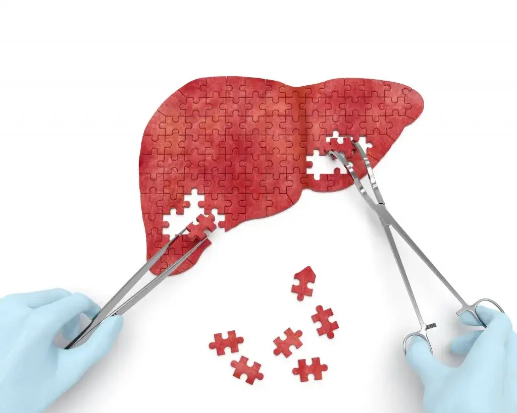
Cholangiocarcinoma
Cholangiocarcinoma is a rare cancer that can develop anywhere within the biliary tree or within the liver. It is commonly identified after the tumor obstructs the bile ducts which results in jaundice and sometimes extreme itching. Cholangiocarcinoma can be located in the lower common bile duct, the mid-portion of the bile duct, where the ducts converge coming out of the liver (Hilar cholangiocarcinoma), or within the liver itself. Hilar cholangiocarcinoma is the most common and also the most complicated to treat given its proximity to important blood vessels entering the liver.
Endoscopic retrograde cholangiopancreatography (ERCP) is a procedure used initially to obtain a biopsy of the tissue, precisely define the location of the tumor and its extent, and to relieve biliary obstruction by the placement of a stent. This type of cancer typically develops a dense inflammatory reaction around it which makes traditional brush biopsies and/or needle biopsies unsuccessful, so a special probe with a camera on the end is placed within the bile duct to visualize the tumor and take direct biopsies with a tiny forceps. Once a biopsy and other tests are performed, a multidisciplinary team including an HPB surgeon, a medical oncologist, a radiation oncologist, and a host of other ancillary support personnel team develops a treatment plan.
Â
Endoscopic retrograde cholangiopancreatography (ERCP)
This procedure is used to obtain a biopsy of the tumor and also to relieve biliary obstruction related to the tumor by the placement of a stent.
Whipple procedure
This procedure is also known as a pancreaticoduodenectomy. It involves removing the head of the pancreas, the duodenum, the distal bile duct, and the gallbladder. This procedure is performed when the tumor is located in the head of the pancreas. The whipple procedure is performed through both open and robotic approaches by Dr. Tierney.
Extrahepatic bile duct resection
This procedure removes the portion of the bile duct that is located between the liver and the head of the pancreas.
Liver resection
This is a surgical operation done to remove a portion of the liver. The liver is the body’s largest internal organ. It plays a vital role in digestion, producing bile to breakdown food.
Before surgery
The steps followed before surgery are:
- An office visit for surgical consultation where a comprehensive history and physical exam is performed.
- Completion of clinical tests for workup and staging.
- Multidisciplinary tumor board review for an expert recommendation regarding treatment strategy.
After surgery
The type of surgical procedure done determines the kind of post-surgery care required. Post care after a surgical operation involves:
- A brief admission to the ICU is sometimes required for complex procedures such as a whipple procedure. Recovery then continues in the hospital ward for another three to five days. Procedures performed robotically typically have a faster recovery and can expect discharge from the hospital one or two days earlier than after open procedures.Â
- The goals of postoperative care include: monitoring for and intervening on any complications that can occur, awaiting the resumption of normal bowel function, maintaining adequate hydration and nutrition, physical and occupational rehabilitation, and providing adequate pain control. Once these measures are met, the patient will be discharged from the hospital. Post-operative follow-up will be scheduled one week from discharge with Dr. Tierney.
Frequently Asked Questions About Cholangiocarcinoma
What is cholangiocarcinoma, and where does it occur?
Cholangiocarcinoma is a cancer that arises from the cells lining the bile ducts. It is rare. Where it sits determines how it behaves and how it is treated, so it is classified by location. Intrahepatic cholangiocarcinoma develops within the liver itself. Hilar cholangiocarcinoma, sometimes called a Klatskin tumour, develops where the ducts converge as they leave the liver, and it is both the most common form and the most technically demanding to treat because it lies immediately alongside the hepatic artery and portal vein. Distal cholangiocarcinoma develops in the lower part of the duct, near the pancreas, and is treated much like a pancreatic head tumour. Risk factors include primary sclerosing cholangitis, choledochal cysts, chronic liver disease and certain parasitic infections, though many people who develop it have no identifiable risk factor.
What are the symptoms of cholangiocarcinoma?
The commonest presentation is painless jaundice, a yellowing of the eyes and skin caused by the tumour obstructing the bile ducts. It is often accompanied by dark urine, pale stools, and itching that can be severe and is frequently what prompts people to seek help. Other symptoms include unexplained weight loss, loss of appetite, fatigue, and discomfort in the upper right abdomen. Fever with jaundice suggests infection in the obstructed ducts and needs urgent assessment. Because the early stages often produce no symptoms at all, the disease is frequently identified only once the duct is blocked, which is one of the reasons it is difficult to treat.
How is cholangiocarcinoma diagnosed?
Diagnosis usually begins with blood tests and imaging, typically a CT scan and an MRCP, a form of MRI that maps the bile ducts in detail. Getting tissue is the harder part. Cholangiocarcinoma provokes a dense inflammatory reaction in the tissue around it, which means conventional brush samples and needle biopsies frequently come back negative even when cancer is present. For that reason a fine camera is passed directly into the bile duct during ERCP so the tumour can be seen and sampled directly with tiny forceps. The same procedure can relieve the obstruction by placing a stent. Once the diagnosis and the extent of disease are established, the case is reviewed by a multidisciplinary team including an HPB surgeon, a medical oncologist, a radiation oncologist and a radiologist, who together plan treatment.
How is cholangiocarcinoma treated?
Surgery offers the only realistic prospect of cure, and whether it is possible depends on where the tumour sits and how far it extends. Tumours within the liver are treated by removing the affected part of the liver. Tumours at the hilum may require removal of the bile ducts together with a section of liver, and reconstruction of the drainage into the intestine. Tumours in the lower duct are usually treated with a Whipple procedure. Because the liver must be left with enough healthy tissue to function, preparation sometimes involves procedures to relieve jaundice or to encourage the remaining liver to grow before surgery. Chemotherapy is used alongside surgery, and where the tumour cannot be removed, treatment focuses on chemotherapy and on relieving the obstruction with stents so that symptoms improve and quality of life is preserved.
What is the outlook with cholangiocarcinoma?
It depends heavily on where the tumour is, how far it has spread, and whether it can be removed completely, and it is worth being honest that the outlook for this cancer is generally poor. Most cases are found at a stage where surgery is not possible, and for those patients treatment aims to control the disease and relieve symptoms rather than to cure. Where the tumour can be completely removed, long-term survival is a genuine possibility, and outcomes are best when the margins are clear and the lymph nodes are not involved. Distal tumours, which are treated like pancreatic head tumours, generally carry a better outlook than hilar ones. These are population patterns and no figure predicts an individual case, which is why the specifics of your imaging and pathology matter far more than any general statistic.